
|

|

|
|

|
|

5. Non-invasive retinal markers may detect people with diabetes and cognitive dysfunction risk
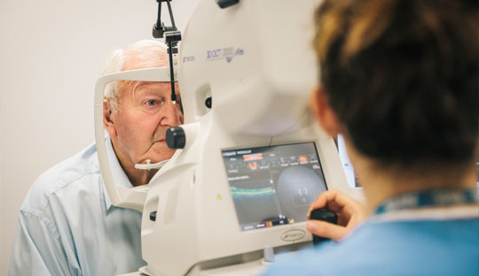
Type 2 diabetes is associated with cognitive impairment and a twofold increased risk of dementia compared to age-matched individuals without diabetes. The eye and the brain share similar embryologic origin and anatomical features and the retina offers a unique “window” to the brain.
The study published in ‘Diabetes’ by a group of researchers headed by Frederik N Pedersen at the Department of Ophthalmology Odense University Hospital determined whether there was any difference in retinal imaging-based neuronal and vascular markers in individuals with type 2 diabetes with or without minimal cognitive impairment (MCI). The cohort included 134 persons with type 2 diabetes. The neuropsychological tests revealed the prevalence of MCI as 28%. 7-field color fundus photos, optical coherence tomography (OCT), OCT-Angiography, and retinal oximetry were performed for the analysis of retinal markers.
The multivariable cluster analysis shows that persons with MCI had significant thinner macular retinal nerve fiber layer and macular ganglion cell layer, and less venular oxygen saturation in the nasal quadrant compared to those without MCI. There were no differences in retinal vessel density, fractal dimension, width, tortuosity or OCT-A markers. People with type 2 diabetes and minimal cognitive impairment demonstrate alterations in retinal structure and metabolism, suggesting that non-invasive retinal markers may be useful to detect people with type 2 diabetes who are at risk of cognitive dysfunction.
For enquiries info@jothydev.net.
Please visit: jothydev.net | research.jothydev.com | diabscreenkerala.net | jothydev.com/newsletter